k A type of bluegreen mold Date of photograph unknown Penicillium on bread. Coloured scanning electron micrograph SEM of the hyphae and fruiting bodies of the fungus Penicillium sp. seen growing as mould on bread. The fruiting bodies, known as conidia, are tuftlike clusters at the tips of the threadlike hyphae. These contain rounded spores strung together in chains. Once ripe each spore can germinate into a new fungus. Species of Penicillium are the source of the antibiotic penicillin. Magnification x194 at 5x7cm size. Magnification x680 at 8x10 ins size. Editorial Stock Photo - Afloimages
Sign up
Login
All images
A type of blue green mold  Date of photograph unknown  Penicillium on bread. Coloured scanning electron micrograph  SEM  of the hyphae and fruiting bodies of the fungus Penicillium sp. seen growing as mould on bread. The fruiting bodies, known as conidia, are tuft like clusters at the tips of the thread like hyphae. These contain rounded spores strung together in chains. Once ripe each spore can germinate into a new fungus. Species of Penicillium are the source of the antibiotic penicillin. Magnification: x194 at 5x7cm size. Magnification  x680 at 8x10 ins size.
ED

A type of blue-green mold (Date of photograph unknown)

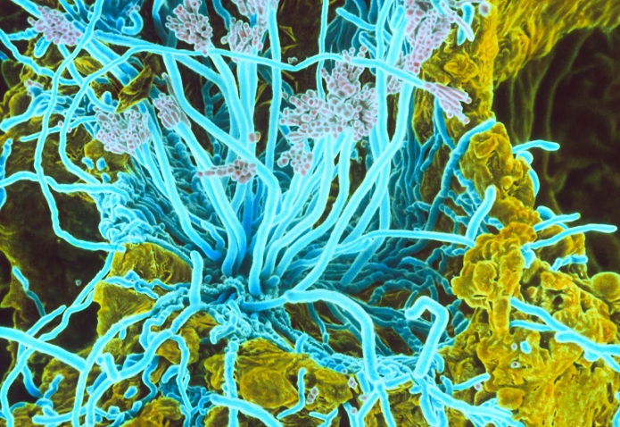
Penicillium on bread. Coloured scanning electron micrograph (SEM) of the hyphae and fruiting bodies of the fungus Penicillium sp. seen growing as mould on bread. The fruiting bodies, known as conidia, are tuft-like clusters at the tips of the thread-like hyphae. These contain rounded spores strung together in chains. Once ripe each spore can germinate into a new fungus. Species of Penicillium are the source of the antibiotic penicillin. Magnification: x194 at 5x7cm size. Magnification; x680 at 8x10 ins size.

Details

ID
10573605

Collection

License type
Editorial

Photographer

Creation date
15-11-2010

Contact Aflo for all commercial uses.


Keywords
More
Sign in
Member access
Login not found.