k A type of bluegreen mold Date of photograph unknown Penicillium fungal spores. Coloured scanning electron micrograph SEM of fruiting bodies of the fungus Penicillium roqueforti. These fruiting bodies conidiophores consist of branching chains of spores conidia, beadlike structures. The mature spores germinate to form new fungi. Penicillium roqueforti is used commercially in the fermentation of blueveined cheeses. Magnification x310 at 6x7cm size. Editorial Stock Photo - Afloimages
Sign up
Login
All images
A type of blue green mold  Date of photograph unknown  Penicillium fungal spores. Coloured scanning electron micrograph  SEM  of fruiting bodies of the fungus Penicillium roqueforti. These fruiting bodies  conidiophores  consist of branching chains of spores  conidia, bead like structures . The mature spores germinate to form new fungi. Penicillium roqueforti is used commercially in the fermentation of blue veined cheeses. Magnification: x310 at 6x7cm size.
ED

A type of blue-green mold (Date of photograph unknown)

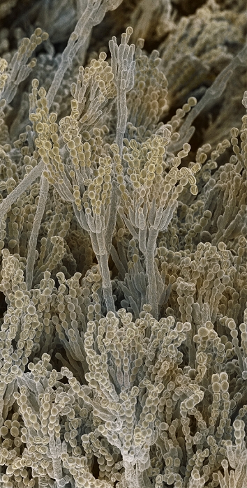
Penicillium fungal spores. Coloured scanning electron micrograph (SEM) of fruiting bodies of the fungus Penicillium roqueforti. These fruiting bodies (conidiophores) consist of branching chains of spores (conidia, bead-like structures). The mature spores germinate to form new fungi. Penicillium roqueforti is used commercially in the fermentation of blue-veined cheeses. Magnification: x310 at 6x7cm size.

Details

ID
10573659

Collection

License type
Editorial

Photographer

Creation date
15-11-2010

Contact Aflo for all commercial uses.


Keywords
More
Sign in
Member access
Login not found.