
ED
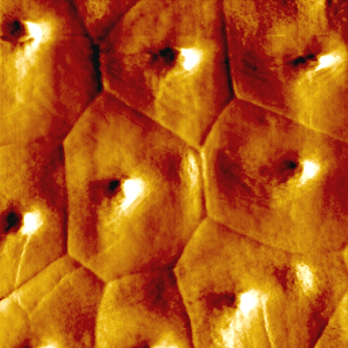
Kidney epithelial cells. Coloured atomic force micrograph (AFM) of the surface of living epithelial cells from the collecting ducts of the kidney. Epithelial cells are flat and line all of the body's surfaces, cavities and lymph and blood vessels. An atomic force microscope is used to study surface at an atomic level. An extremely fine spring-mounted probe is moved across the surface at a constant height. Any deflections are recorded and converted into a computer map of the surface. Magnification: x2000 when printed at 10 centimetres wide.
Details
ID
10610166
Collection
License type
Editorial
Photographer
Creation date
18-11-2010
Contact Aflo for all commercial uses.

More
Top Categories