k Plasmodium falciparum, TEM Plasmodium falciparum plasmodial schizont, or segmenter, in an erythrocyte red blood cell after completion of division, coloured transmission electron micrograph TEM. A residual body is left over after division yellowgreen. The red blood cell has lysed and only a ghost red blood cell membrane no cytoplasm is seen surrounding the new merozoites that are just being released. Free merozoites can be seen outside the ghost red blood cell membrane. The cytoplasm of the merozoites contains a nucleus blue, mitochondria pink and rhoptry bulbs red. Malaria is caused by Plasmodium spp., protozoa. It is spread to humans by Anopheles species mosquitoes. The plasmodial parasite reproduces asexually in red blood cells significantly destroying many of them. Release of mature Plasmodium merozoites results in further infection and produces bouts of shivering fever paroxysms and sweating that may be fatal. Magnification x2,810 when shortest axis Stock Photo - Afloimages
Sign up
Login
All images
Plasmodium falciparum, TEM Plasmodium falciparum plasmodial schizont, or segmenter, in an erythrocyte  red blood cell  after completion of division, coloured transmission electron micrograph  TEM . A residual body is left over after division  yellow green . The red blood cell has lysed and only a ghost red blood cell membrane  no cytoplasm  is seen surrounding the new merozoites that are just being released. Free merozoites can be seen outside the ghost red blood cell membrane. The cytoplasm of the merozoites contains a nucleus  blue , mitochondria  pink  and rhoptry bulbs  red . Malaria is caused by Plasmodium spp., protozoa. It is spread to humans by Anopheles species mosquitoes. The plasmodial parasite reproduces asexually in red blood cells significantly destroying many of them. Release of mature Plasmodium merozoites results in further infection and produces bouts of shivering fever  paroxysms  and sweating that may be fatal. Magnification: x2,810 when shortest axis
RM

Plasmodium falciparum, TEM

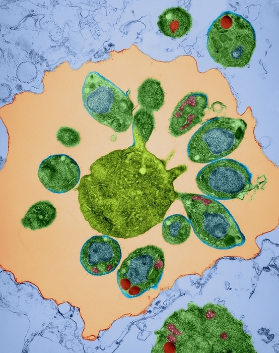
Plasmodium falciparum plasmodial schizont, or segmenter, in an erythrocyte (red blood cell) after completion of division, coloured transmission electron micrograph (TEM). A residual body is left over after division (yellow-green). The red blood cell has lysed and only a ghost red blood cell membrane (no cytoplasm) is seen surrounding the new merozoites that are just being released. Free merozoites can be seen outside the ghost red blood cell membrane. The cytoplasm of the merozoites contains a nucleus (blue), mitochondria (pink) and rhoptry bulbs (red). Malaria is caused by Plasmodium spp., protozoa. It is spread to humans by Anopheles species mosquitoes. The plasmodial parasite reproduces asexually in red blood cells significantly destroying many of them. Release of mature Plasmodium merozoites results in further infection and produces bouts of shivering fever (paroxysms) and sweating that may be fatal. Magnification: x2,810 when shortest axis

Details

ID
153743900

Collection

License type
Rights Managed

Photographer


Sign in
Member access
Login not found.