
RM
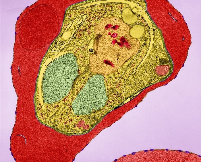
Plasmodium falciparum, TEM
Plasmodium falciparum plasmodial young schizont infecting an erythrocyte (red blood cell), coloured transmission electron micrograph (TEM). The red blood cell membrane has small distinct knobs (purple) that are characteristic of the infection by certain strains of Plasmodium falciparum. The young schizont stage cytoplasm has a distinct food vacuole (light brown) with hemozoin pigment granules (red). The cytoplasm contains two nuclei (ivory), mitochondria (pink) and ribosomes (orange). Maurer's clefts (blue) can be seen in the haemoglobin-containing cytoplasm. Malaria is caused by Plasmodium spp., protozoa. It is spread to humans by Anopheles species mosquitoes. The plasmodial parasite reproduces asexually in red blood cells significantly destroying many of them. Release of mature Plasmodium merozoites results in further infection and produces bouts of shivering fever (paroxysms) and sweating that may be fatal. Magnification: x3,810 when shortest axis

More
Top Categories