
RM
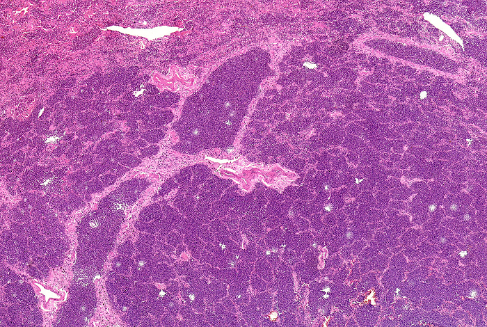
Lobar pneumonia grey hepatisation, light micrograph
Light micrograph section through human lung tissue showing lobar pneumonia grey hepatisation. Grey hepatisation occurs 2 to 3 days following red hepatisation. The lung appears grey with liver-like consistency due to fibrinopurulent exudate, progressive disintegration of red blood cells and hemosiderin. This disease is also known as non-segmental pneumonia or focal non-segmental pneumonia. The most common causes are infections due to bacteria, such as, Streptococcus pneumoniae, Klebsiella pneumoniae and Legionella pneumophila., Photo by NIGEL DOWNER/SCIENCE PHOTO LIBRARY

More
Top Categories