
RM
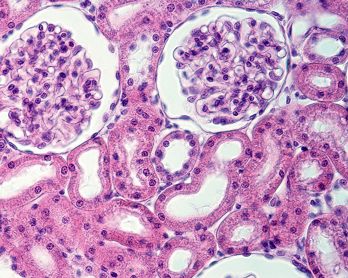
Kidney parenchyma, light micrograph
Light micrograph of the cortex of a kidney fixed by vascular perfusion. The renal parenchyma contains two parts of the nephron, the renal corpuscle and the convoluted tubules. Seen here are two renal corpuscles in which the two main components can be clearly distinguished: the Bowman's capsule, formed by a simple squamous epithelium that surrounds the uriniferous or Bowman's space (white space), and the capillary tuft. The convoluted tubules seen here are all proximal tubules except for two distal convoluted tubules, one of which is at the very centre of the image. In the proximal convoluted tubules the brush border is clearly visible around the lumen. The distal tubules lack a brush border and also have smaller cells (see number of nuclei compared to proximal tubes)., by JOSE CALVO / SCIENCE PHOTO LIBRARY

More
Top Categories