
RM
Typhoid fever, illustration
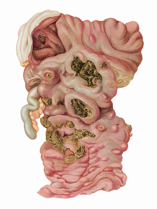
Typhoid fever, illustration. Infection with Salmonella typhi showing swollen mucosa of the ileum with numerous ulcers containing greenish coloured necrotic tissue. The larger ulcers correspond to infected Peyer's patches, the smaller ones from solitary lymphoid follicles. Above, the caecum also displays ulceration. S. typhi is acquired from infected water and food. It invades the gut mucosa and lymphoid tissue, causing ulceration, necrosis and frequent bleeding with possible perforation producing infective peritonitis. If untreated, death is not uncommon. Early antibiotic treatment is effective as a cure. From Bollinger, O. 1901 Atlas und Gundriss der Pathologischen Anatomie, vol 1. Lehmann, Munich., by MICROSCAPE/SCIENCE PHOTO LIBRARY

More
Top Categories