
RM
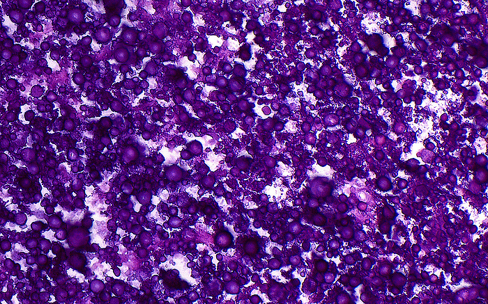
Calcium phosphate calcifications, light micrograph
Light micrograph of calcium phosphate type calcifications. The calcifications, filling the field of view in this image, stain dark purple. Ways these calcifications can form in human tissues include following injury (dystrophic calcification) or due to high levels of calcium in the blood (metastatic calcification). Haematoxylin and eosin stained tissue section. Magnification: 400x when printed at 10cm., by ZIAD M. EL-ZAATARI/SCIENCE PHOTO LIBRARY

More
Top Categories