
RM
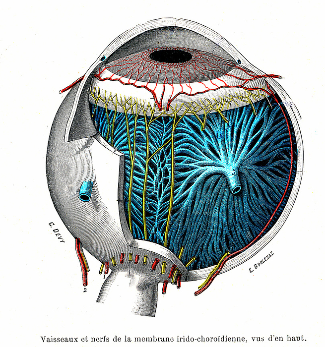
Eye anatomy, illustration
Illustration of the anatomy of the eye. At the front (top) of the eye is the cornea, a transparent coating. Behind this are the pupil and iris. The iris is a coloured muscular ring that regulates the amount of light that enters the pupil. The long posterior ciliary arteries (red, from bottom) and the anterior ciliary arteries (red, around iris) supply blood to the muscles of the iris. Lining the interior of the back of the eye is the retina, the light sensitive membrane. Nerves (blue) transmit the information from the retina to the optic nerve (large nerve at centre right). From Traite d'Anatomie Humaine (1930) by French anatomist Leo Testut (1849-1925). For a labelled version of this image see C057/2788., by COLLECTION ABECASIS/SCIENCE PHOTO LIBRARY

More
Top Categories