
RF
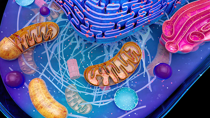
Animal cell, illustration
Illustration of the structure of an animal cell. At top is the endoplasmic reticulum (ER). Some parts of the ER are studded with ribosomes (dots), the cell's protein-manufacturing organelles. Also featured is the Golgi body (pink), associated with the storage and subsequent transport of proteins produced by the ER and mitochondria (brown), the sites of energy synthesis within the cell., by JULIEN TROMEUR/SCIENCE PHOTO LIBRARY
S
0.3 MB
724 x 407 px
6.1 x 3.4 cm
$ 100.00
M
2.8 MB
2290 x 1288 px
19.4 x 10.9 cm
$ 180.00
L
31.6 MB
7680 x 4320 px
65 x 36.6 cm
$ 350.00
More
Top Categories