
RM
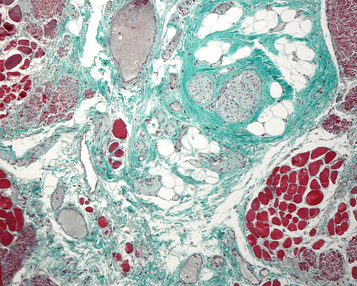
Connective tissue, light micrograph
Light micrograph of connective tissue from the submucosa of the oesophagus wall showing, in the centre, adipocytes, several fascicles of nerve fibres, and small blood vessels (mostly veins) surrounded by collagen fibres stained green by the Masson trichrome method., by JOSE CALVO / SCIENCE PHOTO LIBRARY

More
Top Categories