
RM
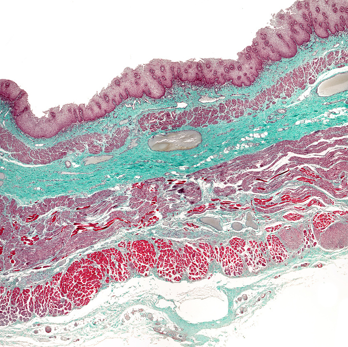
Human oesophagus, light micrograph
Light micrograph of a human oesophagus showing, from top, mucosa lined by a stratified squamous epithelium, lamina propria, muscularis mucosae, submucosa, two muscular layers and adventitia. With Masson trichrome stain, the connective (green) and muscular tissue (light purple) differentiate well., by JOSE CALVO / SCIENCE PHOTO LIBRARY

More
Top Categories