
RM
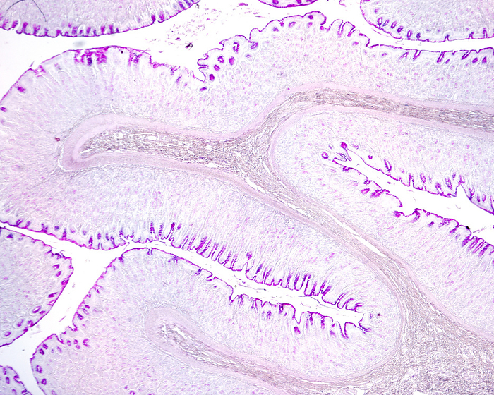
Gastric mucosa, light micrograph
Light micrograph of the gastric wall stained with the Periodic acid-Schiff (PAS) method. The inner layer is the mucosa that shows many folds. In the mucosa, the mucous surface epithelium and foveolar cells of gastric pits show a great PAS positivity. By JOSE CALVO / SCIENCE PHOTO LIBRARY

More
Top Categories