
RM
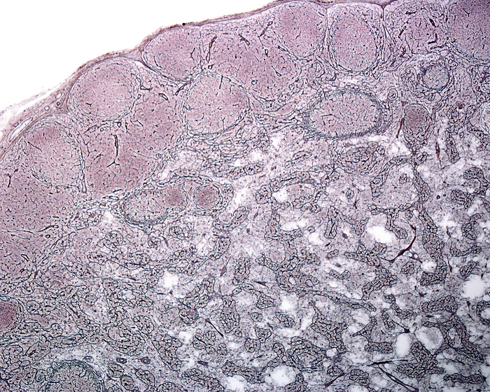
Lymph node reticular fibres, light micrograph
Light micrograph of a lymph node stained with a silver technique for reticular fibres. The capsule and trabeculae appear as thin black lines. The reticular meshwork of reticular fibres can be seen in the lymph node cortex and medulla. The round clear spaces of the cortex (top) are the germinal centres of secondary follicles., by JOSE CALVO / SCIENCE PHOTO LIBRARY

More
Top Categories