
RM
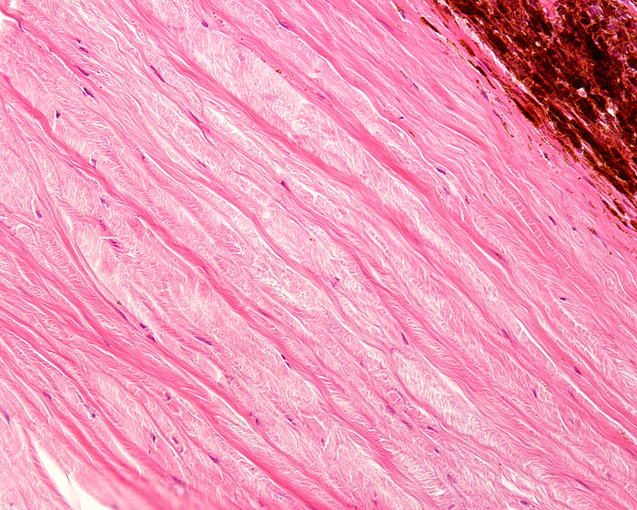
Sclera and lamina fusca, light micrograph
Light micrograph showing the limit between the sclera, white of the eye, and lamina fusca (top right). The sclera is a dense connective tissue made of mainly type I collagen fibres, oriented in different directions. The lamina fusca shows many pigmented cells., by JOSE CALVO / SCIENCE PHOTO LIBRARY

More
Top Categories