
RM
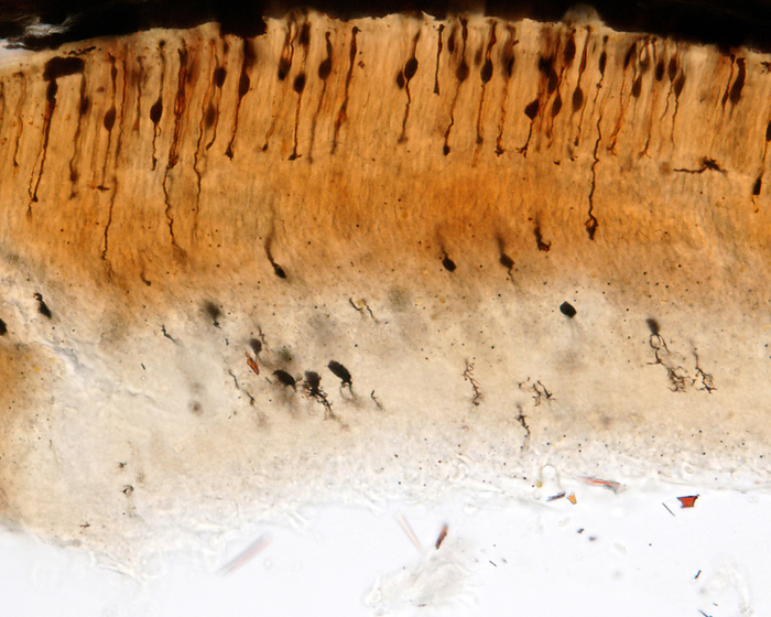
Retina, light micrograph
Light micrograph of a retina stained with the Golgi method. The cells at top are photoreceptors, mainly rods. They show an ovoid-shaped cell body, an upward outer segment and a downward inner segment ending on a rod spherule that synapses with bipolar and horizontal cells., by JOSE CALVO / SCIENCE PHOTO LIBRARY

More
Top Categories