
RM
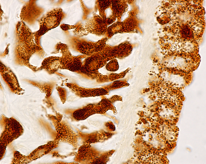
Iris melanin, light micrograph
Light micrograph of the rear surface of an unstained iris showing the localisation of pigment cells. The melanin pigment is concentrated in the rear surface of the iris stroma, and the pigment epithelium (right). Between them there is a clear band that corresponds to the iris dilator muscle., by JOSE CALVO / SCIENCE PHOTO LIBRARY

More
Top Categories