
RM
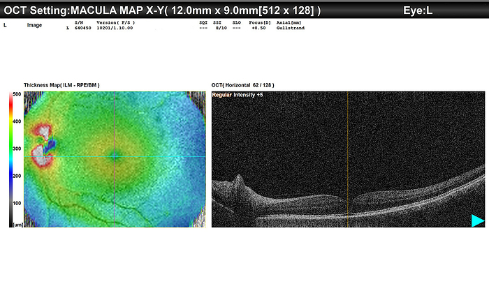
Healthy eye, OCT scan
Optical coherence tomography (OCT) scan (right) and retinal thickness map (left) of a healthy left eye in a 63 year old female patient. The warmer colours are indicative of increased retinal thickness, while cooler colours represent thinner areas. The normal OCT scan shows layers of the retina, alternating in light and dark bands. For scans of the right eye of this patient see SPL code C059/5579., by DR P. MARAZZI/SCIENCE PHOTO LIBRARY

More
Top Categories