
RM
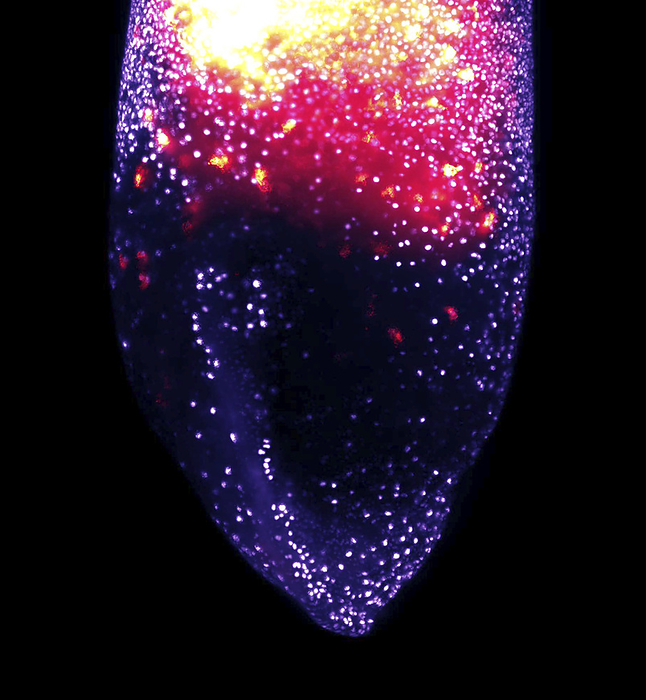
Macrophage development, lattice light-sheet micrograph
Lattice light-sheet micrograph showing the normal development of a population of embryonic macrophages inside a living African clawed frog embryo (Xenopus laevis). The purple dots are the nuclei of the outer cell layer of the embryo (equivalent to the skin) and are labelled with a fluorescent protein. The red cells are the embryonic macrophages underneath the skin layer, visualised with a different fluorescent dye. During normal development, the cells move from the top (near the head region) towards the tail and the two sides of the embryo to distribute throughout the embryo. Macrophages are cells of the immune system. They arise early during embryogenesis and colonise all developing tissues. They phagocytose (engulf) and destroy pathogens, dead cells and cellular debris. See K013/0688 for video clip of this image., by THEBIOCOSMOS/SCIENCE PHOTO LIBRARY

More
Top Categories