
RM
Embryonic macrophages, confocal light micrograph
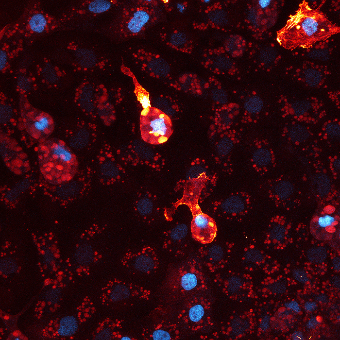
Confocal light micrograph of two macrophages in the precursor to skin tissue in an African clawed frog embryo (Xenopus laevis). The cells are stained to show their cytoskeleton(red) and nuclei (blue). Macrophages are cells of the immune system. They arise early during embryogenesis and colonise all developing tissues. They phagocytose (engulf) and destroy pathogens, dead cells and cellular debris. Magnification: x200 at a printed image size of 10cm., by THEBIOCOSMOS/SCIENCE PHOTO LIBRARY

More
Top Categories