
RM
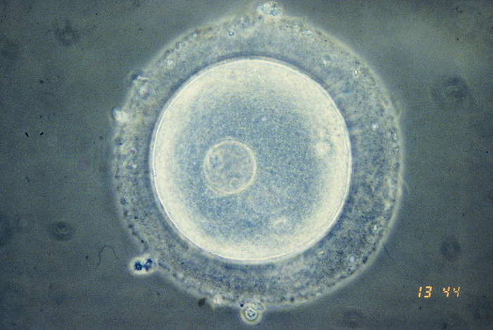
Immature human oocyte, light micrograph
Light micrograph of an immature human oocyte (the female egg cell) containing a germinal body, the large cell nucleus. Oocytes are formed in the ovaries and undergo two meiotic divisions before attaining maturity. Primary oocytes develop in the fetal ovary; further development is arrested at birth & completion of the first meiotic division suspended until after sexual maturity. Fertilisation stimulates the completion of the second meiotic division with the formation of a mature ovum. Magnification: x385 when printed at 10 centimetres wide.

More
Top Categories